Научные публикации
Иконка виртуального микроскопа означает наличие виртуального или видео контента.
Иконка  означает наличие дополнительного статичного иллюстративного материала.
Цветная иконка означает, что материал доступен, тогда как серая  - ожидается.
XXIV Российская конференция по электронной микроскопии (РКЭМ-2012).

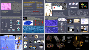
Объемная ультраструктура дендритных синапсов
идентифицированных темных и светлых нейронов гиппокампа крыс.

Работа посвящена сравнительному анализу синапсов темных нейронов, число которых увеличивается при любой патологии мозга.
Полученные данные позволяют говорить о том, что темные нейроны представляют собой нейроны в особом состоянии защиты от
неспецифического возбуждения и эксайтотоксичности.
Темные или гиперхромные нейроны – нейроны с высокой оптической плотностью, выявляемые после прокрашивания или
контрастирования ткани мозга. Природа красителя не имеет значения, это и стандартные красители для гистологии,
и аргирофильные красители для импрегнации серебром по методу Гольджи, и стандартные контрастеры в
электронной микроскопии. Причем темные нейроны можно наблюдать как на окрашенных, так и не окрашенных препаратах.
Одним из первых был Тёрнер, который в 1903 г. описал темные нейроны после механического сдавливания нефиксированных
препаратов спинномозговых ганглиев. С тех пор продолжаются дебаты о том, является ли наблюдаемый феномен признаком
патологии или это артефакт неадекватной химической фиксации и препарирования нервной ткани.
Но практически до 80-х годов никто не рассматривал темные нейроны во взаимосвязи с уровнем их электрической активности,
т.е. как некое функциональное состояние. Даринский провел серию электрофизиологических экспериментов на двигательных
нейронах спинных ганглиев лягушки, в которых убедительно показал, что интенсивность прокрашивания нейронов находится в
прямой зависимости от уровня предшествующей активности этих нейронов, при этом интенсивность прокрашивания нейронов была обратно
пропорциональна глубине наркотизации животного.
В подавляющей массе литературы темные нейроны используют в качестве маркера и критерия выраженности патологии,
а сам феномен связывают с отложенной гибелью этих нейронов. При этом в литературе есть свидетельства о том,
что появление темных нейронов может быть обратимым. В середине 90-х годов группой японских исследователей (под рук. Murakami)
было показано, что темные нейроны в онтогенезе не обнаруживаются, пока не установлены основные нейронные связи и не
сформирован синаптический аппарат. А у взрослых животных наблюдается связь количества темных нейронов и циркадных
ритмов; утром таких нейронов мало, поздно вечером – много. Мы так же наблюдаем массу темных нейронов в ходе
естественной гибернации сусликов, при входе в состояния холодового оцепенения таких нейронов меньше, чем при
выходе из гипотермии, а выход сопровождается взрывной электрической активностью мозга, гипогликемией, оксидативным
стрессом. Суслики за период гибернации проходят до десятка таких циклов входа и пробуждения и если бы все темные нейроны
погибали, то суслик не дожил бы до весны.
Кроме того, масса работ говорит о том, что общая неспецифическая активация синаптической передачи приводит к увеличению
числа темных нейронов и их отложенной гибели, и наоборот, усиление тормозной активности или блокада возбуждающей синаптической
передачи нейроны от гибели спасает.
Нейроны ЦНС – это специализированные клетки, функция которых заключается в обработке информации поступающей
от внутренних органов и из окружающей среды. Элементарными единицами в обработке этой информации выступают
специализированные межклеточные контакты – синапсы, которые, обладая высокой структурно-функциональной пластичностью,
способны менять эффективность передачи сигнала от нейрона к нейрону. Поэтому именно структура синапсов является
отражением функционального состояние нейрона в целом. Однако до сих пор с точки зрения синаптической пластичности
темные нейроны никто не рассматривал.
Еще десятилетие назад Engert и Bonhoeffer в Nature (1999) показали, что для появления новых синапсов
in vitro требуется несколько десятков минут, а in vivo этот процесс занимает еще больше времени,
тогда как химическая фиксации структуры ткани инициируется в считанные секунды. Наша идея заключалась в том, что если темный
нейрон - это артефакт препарирования и
неадекватной фиксации ткани, то функциональная структура его синапсов не должна отличаться от структуры
синапсов светлых нейронов. Если же она отличается, то это говорит как раз об обратном, т.е. темный нейрон
представляет собой особое функциональное состояние. Именно эту идею мы и попытались проверить в нашем пилотном исследовании
с использованием методов объемной (3D) реконструкции.
Дополнительные иллюстрации к слайдам 8-10
 ,
и видеоматериалы к
 слайду 5,
 слайду 10,
 слайду 12.
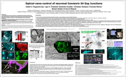
Optical nano-control of neuronal Connexin-36 Gap Junctions
Four lines of evidence support the idea that neuronal Gap Junctions (GJ) are operating in accord with chemical synaptic transmission:
I. First, electron microscopy analyses of brain sections revealed typical double membrane of GJ that are often present close to the synapses
(our own data:  Suppl. Fig.3.;
 Suppl. Fig.6.;
 Suppl. video).
II. Second, the amplitude of neural responses in many brain regions found to be modified. This fact would be difficult to explain without appreciation
of electrical synapses and Belousov's group showed that NMDA receptors regulate developmental GJ uncoupling via CREB signaling.
III. Cx36-deficient mice demonstrate that transmission through electrical synapses is important for neuron and brain function. Generation and analyses of Cx36-GFP expressing mice revealed that electrical synapses are abundant in the mice brain and
their function believed to be important for the generation of synchronous oscillations. The functional consequences of electrical
synapses are still incompletely understood, but recent reports documented abnormal circadian activity, deficits in motor-coordination, motor learning, and impaired memory recall.
IV. Biochemical analyses, show that Cx36 present in a complex with the scaffold protein zonula occludens (ZO-1).
Thus, neuronal coupling via GJs is extremely important in early development. Studies of GJ coupling between interneurons in the cortex, amygdala, and hippocampus, shown to be mediated mainly by
Cx36, although in some instances electrical synapses may include other connexins as e.g. Cx45, Cx47, Cx57. It still remained to be demonstrated
that considerable specificity in connexin distribution in brain play an important role in electrically-coupled neural circuits.
Objectives:
• to understand how neuronal connexins are maintained in the plasma membrane we performed proteomics screened from brain extracts in search for molecules interacting
with cytosolic moieties of Connexin 36 and preferentially localized to the plasma membrane.
• to test whether found by proteomics molecules have biological relevance we analysed their effects on Cx36 in living cells.
Methods: Electron microscopy (EM), Cryo-EM, Proteomics, Nano-Spectroscopy, and Live cell imaging:
High Resolution Spinning Disk Nikon based set-up with CO2, z-PFS, anisiotropy, FCS and
dual split FRET measurements devices were set to resolve cellular structures and protein-protein
interactions in living cells.
Results: I. We demonstrate here that newly described protein Drebrin (Developmentally REgulated BRain proteIN) may directly interact with Cx36 in living cells and removal of drebrlin may have consequences on the stability and formation of neuronal cell-cell contacts.
II. Second, we show that cells may store connexins in the ER under unfavorable conditions (e.g.  Suppl. Fig.5.). If the activity-dependent transport of connexins to the PM is delayed, Cx36may undergo ER associated degradation.
III. Mapping Cx36 domains and testing them against corresponding domains of Drebrin revealed potential sites in Cx36 cytosolic loop and tail that may have biological relevance for in vivo function and thereby incorporation of Cx36 channels into zones adjacent to electrical synapses.
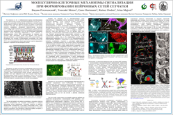
Molecular and cellular mechanisms of signaling in Retina:
Lessons from the developmental regulation of neuronal circuit formation
Recently we have shown that newly formed horizontal cells (HC) and amacrine cells (AC) layers express high level of Drebrin - Developmental Regulated Brain Protein.
We showed direct binding of Cx43 to drebrin that was analysed in details in previous work. Here we perform in vivo and in vitro analyses for the role of drebrin,
that expressed in two splice isoformes E and A - Adult and Embryonic). Our idea was that drebrin may be required for connexin signaling during fast rearrangements
of cell-cell contacts and upon establishment of retinal layer specificity. Both HC and AC are also known to express array of neuronal connexins, Cx36, Cx45, Cx57.
The functional link between Drebrin and Connexins remained obscur. Gap junctions (GJ) appear to be involved in restricts tracer coupling between neighboring HC
in newly formed layer of retina and between AC but not yet in ganglion-ganglion cell coupling. However, tracer coupling between HC and amacrine cells at the earliest
ages is yet to be defined, probably to the rearrangements of cell-cell contacts during developmental migration. At this time (E15-20), combinations of glutamate
antagonists or GABA-A antagonist does not influence cell cell-coupling. The direct roles of connexins in HC and AC layers is still remained to be analysed in greater
details. One idea is that GJ coupling between HC and ACs (possibly the cholinergic ACs) is a transient early phase of transmission
employed before the chemical synapses.
Interestingly, completely mature electrical transmission and generation of retinal waves in mammals can proceed via GJ before synaptic.
Connexins are known to be permeable to cAMP. This second messenger can modulate intracellular levels of cAMP in a cluster of connected cells and have
dramatic effects on the propagation of Ca2+ waves. Thus, GJs play a very important role in the cell-layer definition before chemical synapses have
started to act.
To better understand the role of early Drebrin expression we combined in utero electroporation of drebrin RNAi in embryo brain with in vitro cellular approaches and
expressed Drebrin together with neuronal connexins in neuronal and non-neuronal cells. Our in vitro reconstruction experiments revealed stunning role of Drebrin in
stabilizing neuronal connexins at the cell-cell interface. To control the obtained data we also applied RNAi of Drebrin and showed that in the absence of drebrin
the neuronal connexins were unable to be delivered to the plasma membrane and to form contacts with neighboring cells. Single cell assay of RNAi using high resolution
Live Cell Microscopy show how in the absence of drebrin Cx36 was unable to be delivered to the plasma membrane and to establish cell-cell. Instead,
unsupported by submembrane cytoskeletal scaffold Cx36 was co-localized with lysosomal markers and revealed degradation bands when analyzed in WB and Cx36 specific antibodies.
The obtained results confirm ability of highly morphogenic protein drebrin to stabilize and spatially regulate rearrangements of cell-cell contacts thus quickly
providing the adjustability of intercellular signaling. This unexpected finding not only adds to the developmental complexity in regulation of signaling but also
shows how cross-talks between cytoskeleton and cell-cell connectivity may influence complex processes as signaling and transmission of visual signal in retina and
brain. Cx36 in known to be the most permeable to the cAMP and broadly expressed in inhibitory GABA-ergic interneurons. Thus drebrin-induced rearrangements of
cell-cell contacts may regulate not only excitatory pathways of retinal PR but also provide complex inhibitory regulation of ganglion cells. The question still
remained to answer, how the expression of drebrin itslelf is regulated in development and under local conditions.
 Дополнительный видеоматериал к Рис.6.
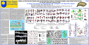
3D реконструкция синапсов зубчатой фасции крысы на основе серийных ультратонких срезов
при долговременной потенциации in vivo.
Считается, что научение и закрепление памяти связанны с изменениями функциональной активности синапсов (силы синапса или его вклада в генерацию постсинаптического ответа), что с другой стороны может быть связано со структурой синапсов вовлеченных в этот процесс. Играет ли роль количество синапсов в единице объема нервной ткани (плотность синапсов), или их структура, а возможно и то и другое, в процессах закрепления памяти в целом малоизученно. Знания о плотности синапсов и таких их ультраструктурных особенностях как объем, площадь поверхности мембраны, форма как постсинаптических уплотнений (PSD) и дендритных шипиков (DS) – очень важные факторы для оценки морфологических коррелятов уровня синаптической активности/эффективности. Долговременная потенциация (LTP – long term potentiation) представляет собой клеточную модель в исследованиях научения и памяти. Считается, что развитие поздней фазы LTP (L-LTP – late-LTP) требует усиления синтеза белка (и in vivo и in vitro) и развивается спустя 2-5 часов после запуска LTP. Поэтому, если какие-либо значительные структурные изменения синапсов и можно было бы наблюдать, то, вероятнее всего, по окончании этого периода.
Для того чтобы понять функциональную роль различных типов дендритных синапсов после индукции LTP мы использовали стереологический анализ и 3D реконструкцию синапсов молекулярного слоя зубчатой фасции гиппокампа на серийных ультратонких срезах (70 нм толщиной), согласно методам разработанным Кристен Харрис. В гиппокампах левого полушария вызывали LTP тетанической стимуляцией перфорирующего пути гиппокампа, а правое полушарие служило контролем.
Согласно классификации дендритных шипиков, предложенной Кайзерман-Абрамовым
(Kaiserman-Abramof, 1969)
на основе импрегнации нейронов коры больших полушарий по методу Гольджи и расширенной классификацией Кристен Харрис на ультраструктурном уровне с использованием 3D реконструкции синапсов гиппокампа
(Harris et al., 1992), мы показали, что в молекулярном слое зубчатой фасции гиппокампа синапсы представлены четырьмя категориями аксо-шипиковых и стволовых синапсов. Три категории из них были классифицированы согласно форме шипиков, на которых они располагались – это синапсы грибовидные
(на грибовидных дендритных шипиках  ),
тонкие (на тонких дендритных шипиках
  ),
пеньковые (на пеньковых дендритных шипиках
  )
и четвертая категория – синапсы стволовые (на стволах дендритов).
 .
.
Although there were no differences in synapse densities in different areas of dentate gyrus molecular sublayers (stereological analysis performed on whole series of sections), we marked reliable changes in distribution of fourth categories of synapses for LTP and control state. Differences were revealed between portion of thin, stubby, and shaft synapses in LTP vs. control. No significant differences for density of mushroom synapses for all states were revealed.
We have performed quantitative analysis of 3D reconstructions of two categories of synapses having expressed DSs with PSDs: thin and mushroom. For analysis, about 100 spines per each state were reconstructed/analyzed. Induction of the LTP induces the increasing of volume and area of thin spines. One-Way ANOVA results in reliable differences between control and LTP. Analysis of PSDs in both control and LTP reveal the increasing also in PSD size during LTP.
Central panels "a" and "b" show the subsets of DSs, randomly selected for control and LTP. "a" – spines located on distal part of granule cell dendrites while "b" – represent the spines from proximal dendrites. Even visual analysis shows that LTP induces the changes in shape of the spine. In LTP the DSs have concave shape in contrast to convex shape in control.
Induction of the LTP also induces the increasing of volume (and area) of thin spines. Statistic analysis shows very reliable differences between control and LTP for both parameters.
In conclusion: data obtained suggest that L-LTP induces sharp transformations in the structure of both thin and mushroom dendritic spine synapses following 6h post-tetanic stimulation of perforant path in vivo, which represent the most prominent effect related to learning and memory formation in comparison to changes in synapse density.


 Презентация, РКЭМ-2012, Черноголовка.
 текст доклада
 текст постера
