Publications
Virtual microscope icon  means that you can see supplemental virtual or video content,
whereas icon with the sign plus  means presence of supplementary static materials.
Color distinct icon means, that data are now available, whereas fade icon  means expected.
FENS Forum 2010
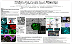
Optical nano-control of neuronal Connexin-36 Gap Junctions
Four lines of evidence support the idea that neuronal Gap Junctions (GJ) are operating in accord with chemical synaptic transmission:
I. First, electron microscopy analyses of brain sections revealed typical double membrane of GJ that are often present close to the synapses
(our own data:  Suppl. Fig.3.;
 Suppl. Fig.6.;
 Suppl. video).
II. Second, the amplitude of neural responses in many brain regions found to be modified. This fact would be difficult to explain without appreciation
of electrical synapses and Belousov's group showed that NMDA receptors regulate developmental GJ uncoupling via CREB signaling.
III. Cx36-deficient mice demonstrate that transmission through electrical synapses is important for neuron and brain function. Generation and analyses of Cx36-GFP expressing mice revealed that electrical synapses are abundant in the mice brain and
their function believed to be important for the generation of synchronous oscillations. The functional consequences of electrical
synapses are still incompletely understood, but recent reports documented abnormal circadian activity, deficits in motor-coordination, motor learning, and impaired memory recall.
IV. Biochemical analyses, show that Cx36 present in a complex with the scaffold protein zonula occludens (ZO-1).
Thus, neuronal coupling via GJs is extremely important in early development. Studies of GJ coupling between interneurons in the cortex, amygdala, and hippocampus, shown to be mediated mainly by
Cx36, although in some instances electrical synapses may include other connexins as e.g. Cx45, Cx47, Cx57. It still remained to be demonstrated
that considerable specificity in connexin distribution in brain play an important role in electrically-coupled neural circuits.
Objectives:
• to understand how neuronal connexins are maintained in the plasma membrane we performed proteomics screened from brain extracts in search for molecules interacting
with cytosolic moieties of Connexin 36 and preferentially localized to the plasma membrane.
• to test whether found by proteomics molecules have biological relevance we analysed their effects on Cx36 in living cells.
Methods: Electron microscopy (EM), Cryo-EM, Proteomics, Nano-Spectroscopy, and Live cell imaging:
High Resolution Spinning Disk Nikon based set-up with CO2, z-PFS, anisiotropy, FCS and
dual split FRET measurements devices were set to resolve cellular structures and protein-protein
interactions in living cells.
Results: I. We demonstrate here that newly described protein Drebrin (Developmentally REgulated BRain proteIN) may directly interact with Cx36 in living cells and removal of drebrlin may have consequences on the stability and formation of neuronal cell-cell contacts.
II. Second, we show that cells may store connexins in the ER under unfavorable conditions (e.g.  Suppl. Fig.5.). If the activity-dependent transport of connexins to the PM is delayed, Cx36may undergo ER associated degradation.
III. Mapping Cx36 domains and testing them against corresponding domains of Drebrin revealed potential sites in Cx36 cytosolic loop and tail that may have biological relevance for in vivo function and thereby incorporation of Cx36 channels into zones adjacent to electrical synapses.
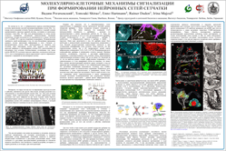
Molecular and cellular mechanisms of signaling in Retina:
Lessons from the developmental regulation of neuronal circuit formation
Recently we have shown that newly formed horizontal cells (HC) and amacrine cells (AC) layers express high level of Drebrin - Developmental Regulated Brain Protein.
We showed direct binding of Cx43 to drebrin that was analysed in details in previous work. Here we perform in vivo and in vitro analyses for the role of drebrin,
that expressed in two splice isoformes E and A - Adult and Embryonic). Our idea was that drebrin may be required for connexin signaling during fast rearrangements
of cell-cell contacts and upon establishment of retinal layer specificity. Both HC and AC are also known to express array of neuronal connexins, Cx36, Cx45, Cx57.
The functional link between Drebrin and Connexins remained obscur. Gap junctions (GJ) appear to be involved in restricts tracer coupling between neighboring HC
in newly formed layer of retina and between AC but not yet in ganglion-ganglion cell coupling. However, tracer coupling between HC and amacrine cells at the earliest
ages is yet to be defined, probably to the rearrangements of cell-cell contacts during developmental migration. At this time (E15-20), combinations of glutamate
antagonists or GABA-A antagonist does not influence cell cell-coupling. The direct roles of connexins in HC and AC layers is still remained to be analysed in greater
details. One idea is that GJ coupling between HC and ACs (possibly the cholinergic ACs) is a transient early phase of transmission
employed before the chemical synapses.
Interestingly, completely mature electrical transmission and generation of retinal waves in mammals can proceed via GJ before synaptic.
Connexins are known to be permeable to cAMP. This second messenger can modulate intracellular levels of cAMP in a cluster of connected cells and have
dramatic effects on the propagation of Ca2+ waves. Thus, GJs play a very important role in the cell-layer definition before chemical synapses have
started to act.
To better understand the role of early Drebrin expression we combined in utero electroporation of drebrin RNAi in embryo brain with in vitro cellular approaches and
expressed Drebrin together with neuronal connexins in neuronal and non-neuronal cells. Our in vitro reconstruction experiments revealed stunning role of Drebrin in
stabilizing neuronal connexins at the cell-cell interface. To control the obtained data we also applied RNAi of Drebrin and showed that in the absence of drebrin
the neuronal connexins were unable to be delivered to the plasma membrane and to form contacts with neighboring cells. Single cell assay of RNAi using high resolution
Live Cell Microscopy show how in the absence of drebrin Cx36 was unable to be delivered to the plasma membrane and to establish cell-cell. Instead,
unsupported by submembrane cytoskeletal scaffold Cx36 was co-localized with lysosomal markers and revealed degradation bands when analyzed in WB and Cx36 specific antibodies.
The obtained results confirm ability of highly morphogenic protein drebrin to stabilize and spatially regulate rearrangements of cell-cell contacts thus quickly
providing the adjustability of intercellular signaling. This unexpected finding not only adds to the developmental complexity in regulation of signaling but also
shows how cross-talks between cytoskeleton and cell-cell connectivity may influence complex processes as signaling and transmission of visual signal in retina and
brain. Cx36 in known to be the most permeable to the cAMP and broadly expressed in inhibitory GABA-ergic interneurons. Thus drebrin-induced rearrangements of
cell-cell contacts may regulate not only excitatory pathways of retinal PR but also provide complex inhibitory regulation of ganglion cells. The question still
remained to answer, how the expression of drebrin itslelf is regulated in development and under local conditions.
 Suppl. video for Fig.6.
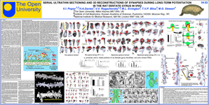
Serial ultrathin sectioning and 3D reconstructions of synapses during LTP
in the rat dentate gyrus in vivo.
It is generally accepted that learning and memory consolidation are related to changes in synaptic strength, which in turn related to structure of the synapses involved. Whether density of synapses, or their structure, or both, are playing role in memory consolidation, generally unknown. Knowledges about the density of synapses and fine details (volume, area, shape) of both postsynaptic densities (PSD) and dendritic spines (DS) are very important in the search of morphological correlates of synaptic transmission. Long-term potentialion (LTP) represent cellular model of learning and memory. Late phase of LTP (L-LTP) requires enhanced protein synthesis (both in vivo and in vitro) is believed to begin 2-5 h after induction of LTP. This argues that more prominent synaptic structural changes, if any, are more likely to occur after this period.
To understand better the functional implication of the different types of synapses after L-LTP induction in the molecular layer (ML) of hippocampal dentate gyrus (DG) both stereological analysis and 3D reconstructions of serial 70nm sections were used according to methodology of K.M. Harris. Hippocampus from left hemisphere was subjected to tetanic electrical stimulation while that of from right hemisphere was used as "control".
Accordingly to Golgi studies of DSs by
Kaiserman-Abramof (1969) in cortex,
and by
Harris et al. (1992) with 3D reconstructions on hippocampus, four categories of axo-spinous and shaft synapses in molecular layer of dentate gyrus were classified as
mushroom spines  ,
thin DSs
  ,
stubby DSs
  ,
and shaft synapses. That is three categories of them were classified accordingly to the shape of spines
 .
.
Although there were no differences in synapse densities in different areas of dentate gyrus molecular sublayers (stereological analysis performed on whole series of sections), we marked reliable changes in distribution of fourth categories of synapses for LTP and control state. Differences were revealed between portion of thin, stubby, and shaft synapses in LTP vs. control. No significant differences for density of mushroom synapses for all states were revealed.
We have performed quantitative analysis of 3D reconstructions of two categories of synapses having expressed DSs with PSDs: thin and mushroom. For analysis, about 100 spines per each state were reconstructed/analyzed. Induction of the LTP induces the increasing of volume and area of thin spines. One-Way ANOVA results in reliable differences between control and LTP. Analysis of PSDs in both control and LTP reveal the increasing also in PSD size during LTP.
Central panels "a" and "b" show the subsets of DSs, randomly selected for control and LTP. "a" – spines located on distal part of granule cell dendrites while "b" – represent the spines from proximal dendrites. Even visual analysis shows that LTP induces the changes in shape of the spine. In LTP the DSs have concave shape in contrast to convex shape in control.
Induction of the LTP also induces the increasing of volume (and area) of thin spines. Statistic analysis shows very reliable differences between control and LTP for both parameters.
In conclusion: data obtained suggest that L-LTP induces sharp transformations in the structure of both thin and mushroom dendritic spine synapses following 6h post-tetanic stimulation of perforant path in vivo, which represent the most prominent effect related to learning and memory formation in comparison to changes in synapse density.


 FENS Forum 2010, Amsterdam.
 Plain text from poster.

